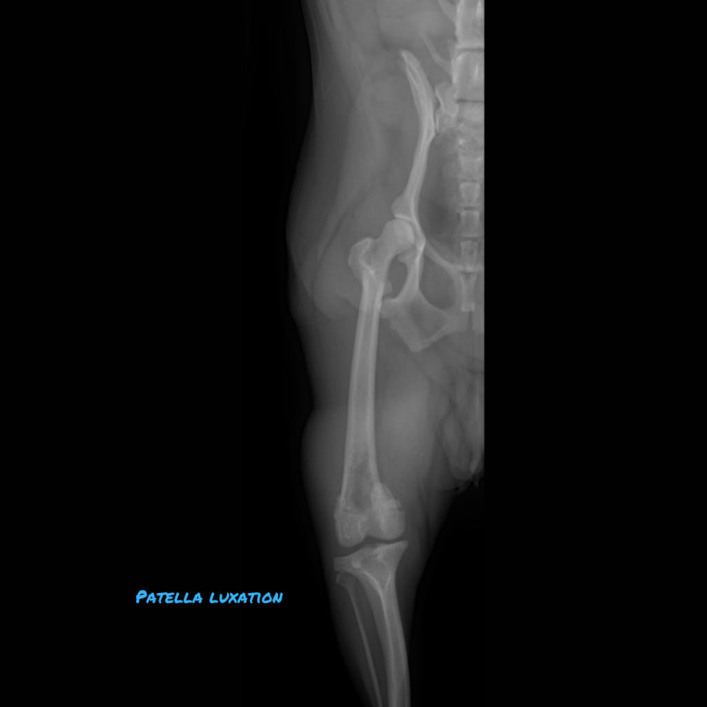
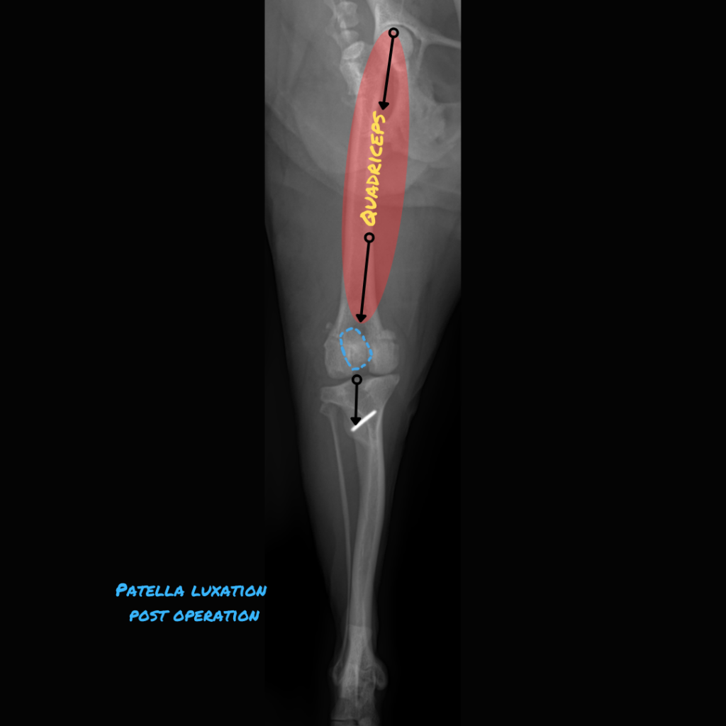
Patellar luxation occurs when the kneecap slips out of its groove, shifting medially or laterally. The patella is embedded within the tendon that connects the quadriceps muscle to the tibial tuberosity, assisting in knee straightening. The patella shields the tendon as it glides over the end of the thigh bone.
Most luxations are medial and caused by anatomic abnormalities, although traumatic luxations can also occur. Anatomic factors can also cause lateral luxation, usually in young animals.
.
Luxation are graded as follows:
Grade 1: Patella can be manually luxated but it immediately returns to a normal position when released.
Grade 2: Patella luxates and returns to a normal position spontaneously. Luxation resolves with the joint flexed. Grade 2 luxation can progress to a grade 3 luxation over time or if concurrent cruciate ligament disease develops.
Grade 3: Patella is usually luxated but can be manually reduced. After manual reduction the patella luxates again immediately once released.
Grade 4: Patella is luxated permanently and cannot be manually replaced into the trochlear groove.
.
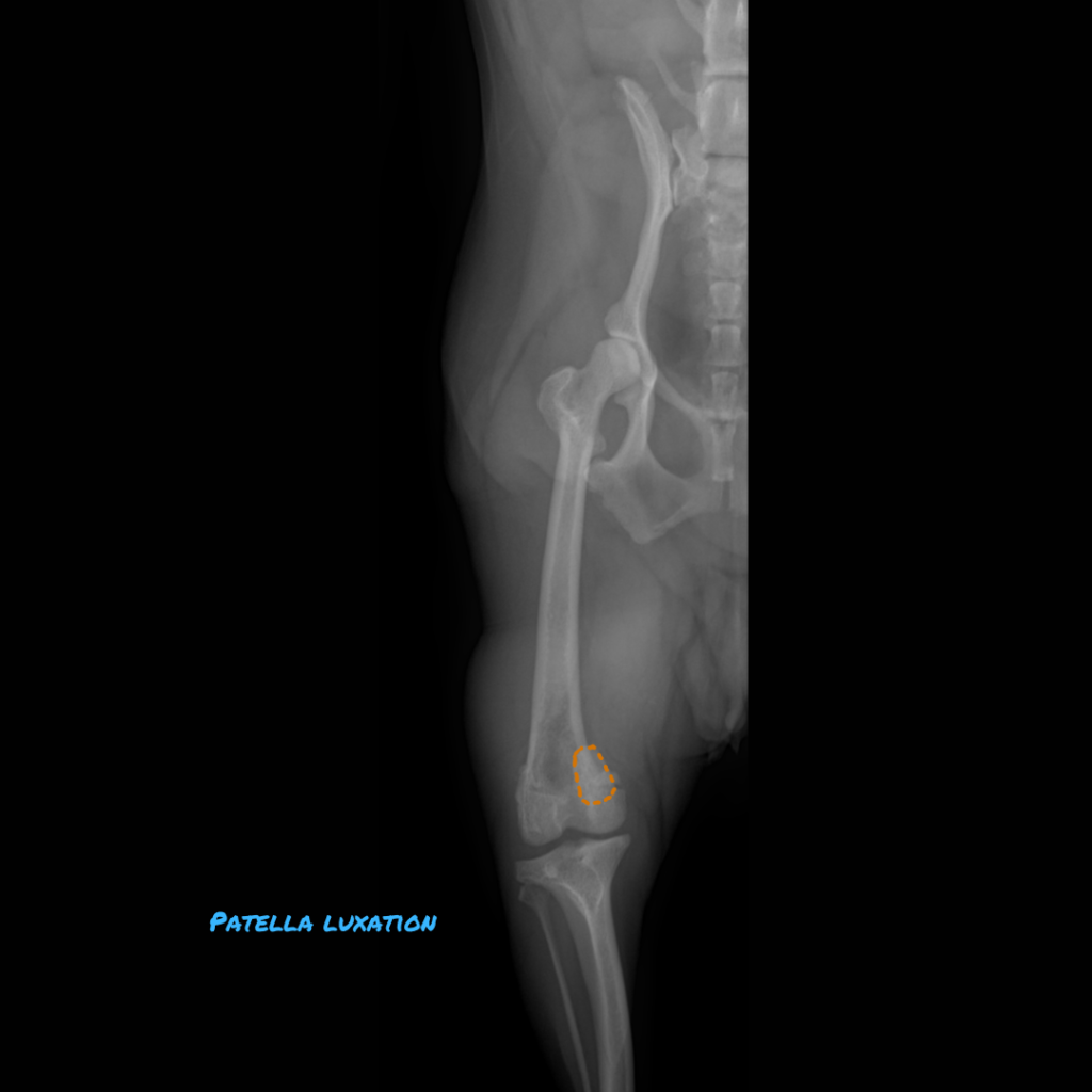
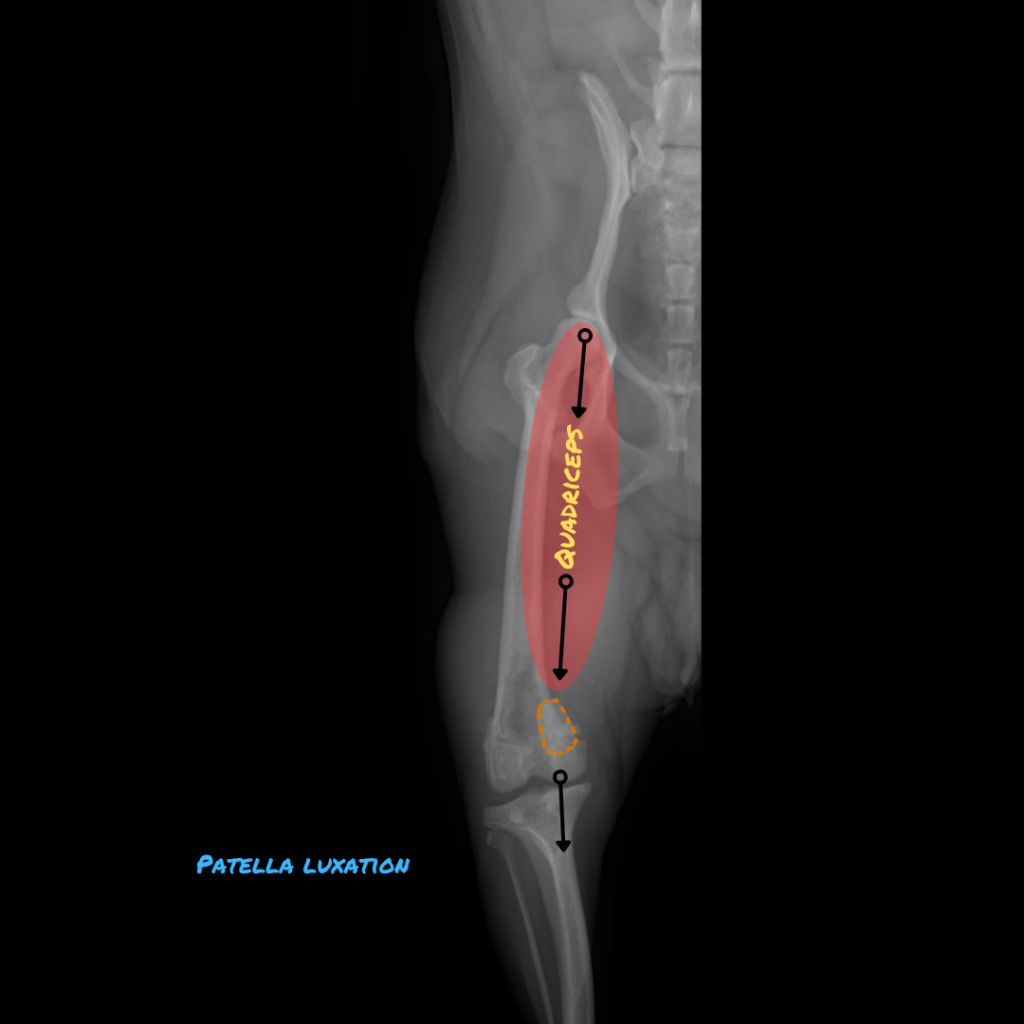
[Pic 1: The patella is highlighted with an orange dotted line and is luxated medially]
.
As a rule of thumb, dogs with clinical signs associated with patellar luxation of any grade may be considered for surgical correction. Such signs may include persistent lameness or multiple, frequent episodes of lameness. Grade 3 and 4 luxations most frequently benefit from surgery, while grade 2 luxations may also benefit if they meet the above criteria.
.
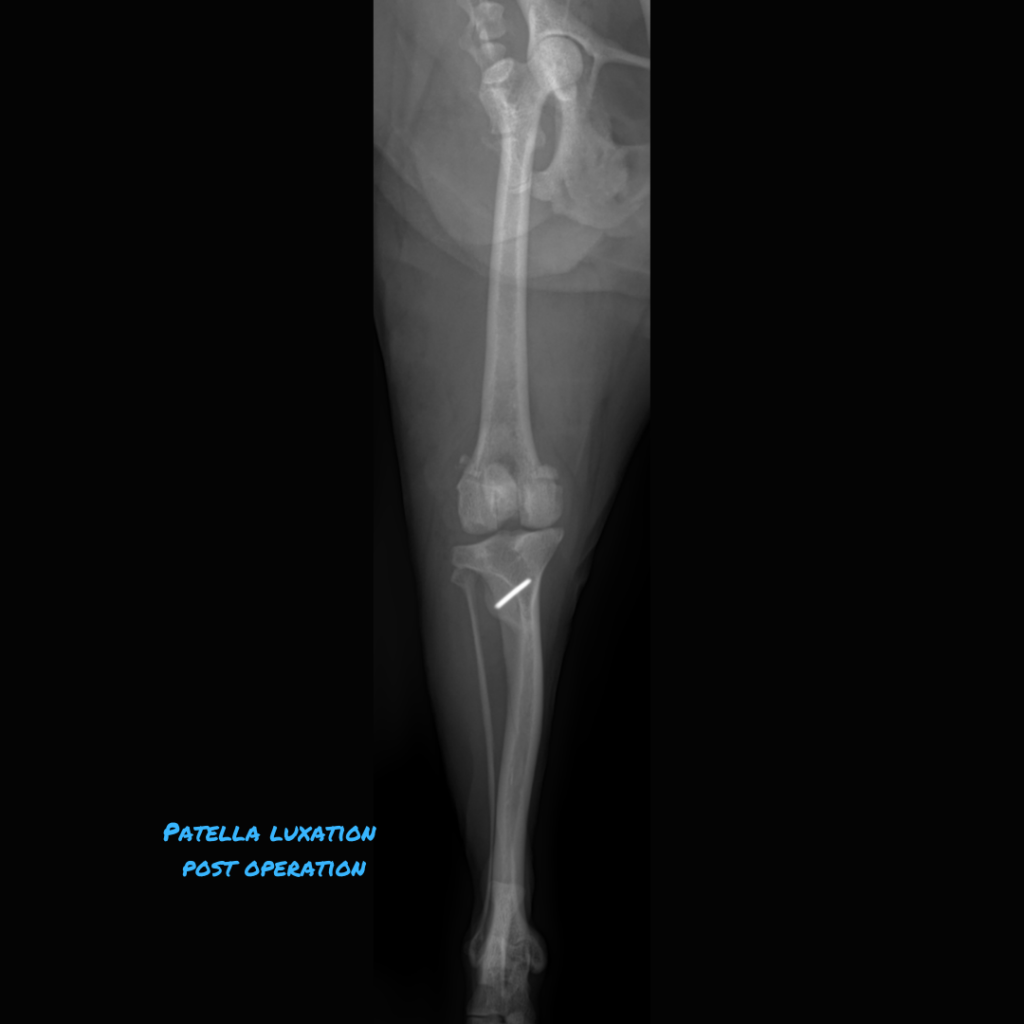
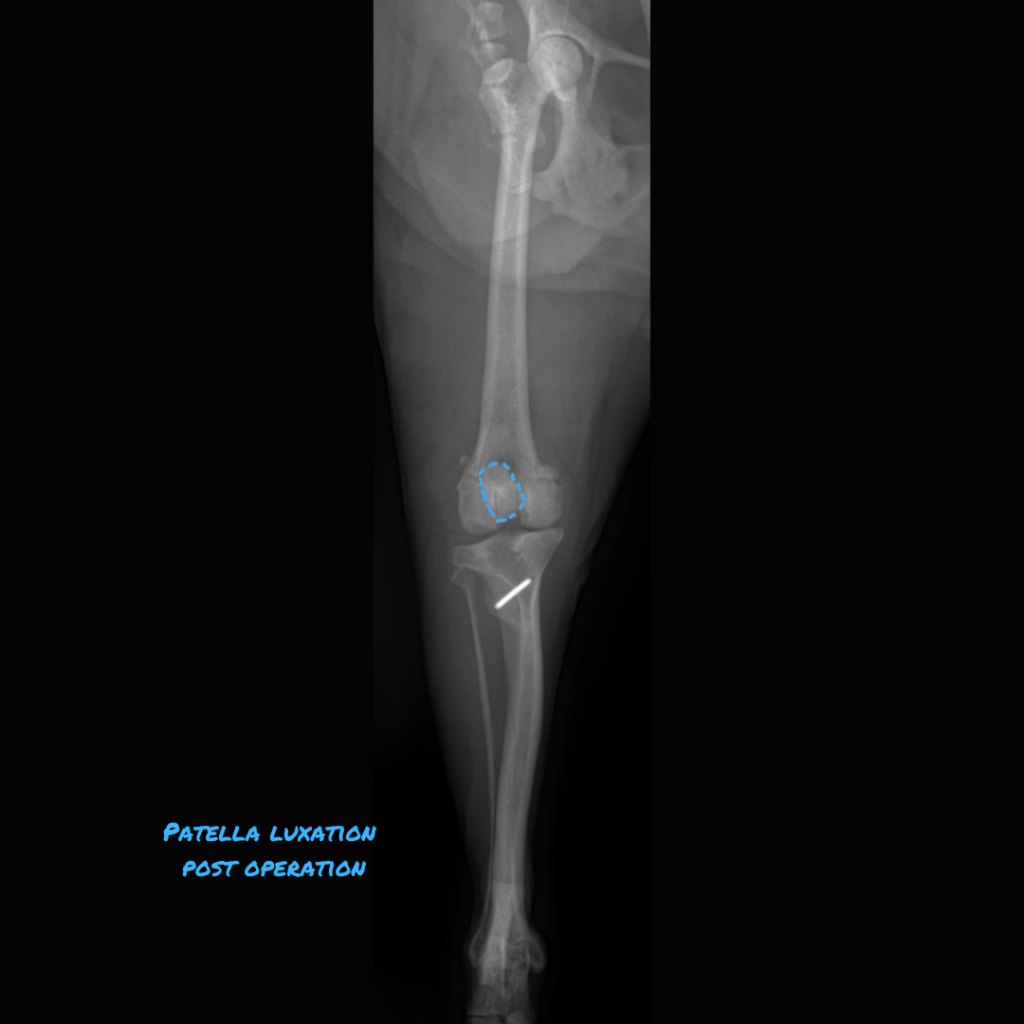
[Pic 2: The tibial tuberosity was shifted laterally and the groove was made deeper, so the patella is now sitting in its proper spot in the middle. (blue dotted line)
.
P.S: It is recommended to consult with your veterinarian regarding the surgical treatment of patella luxation