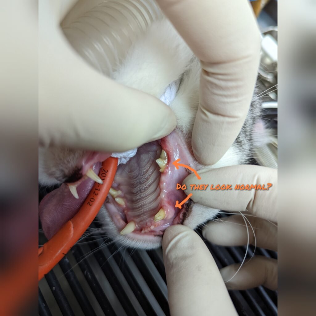
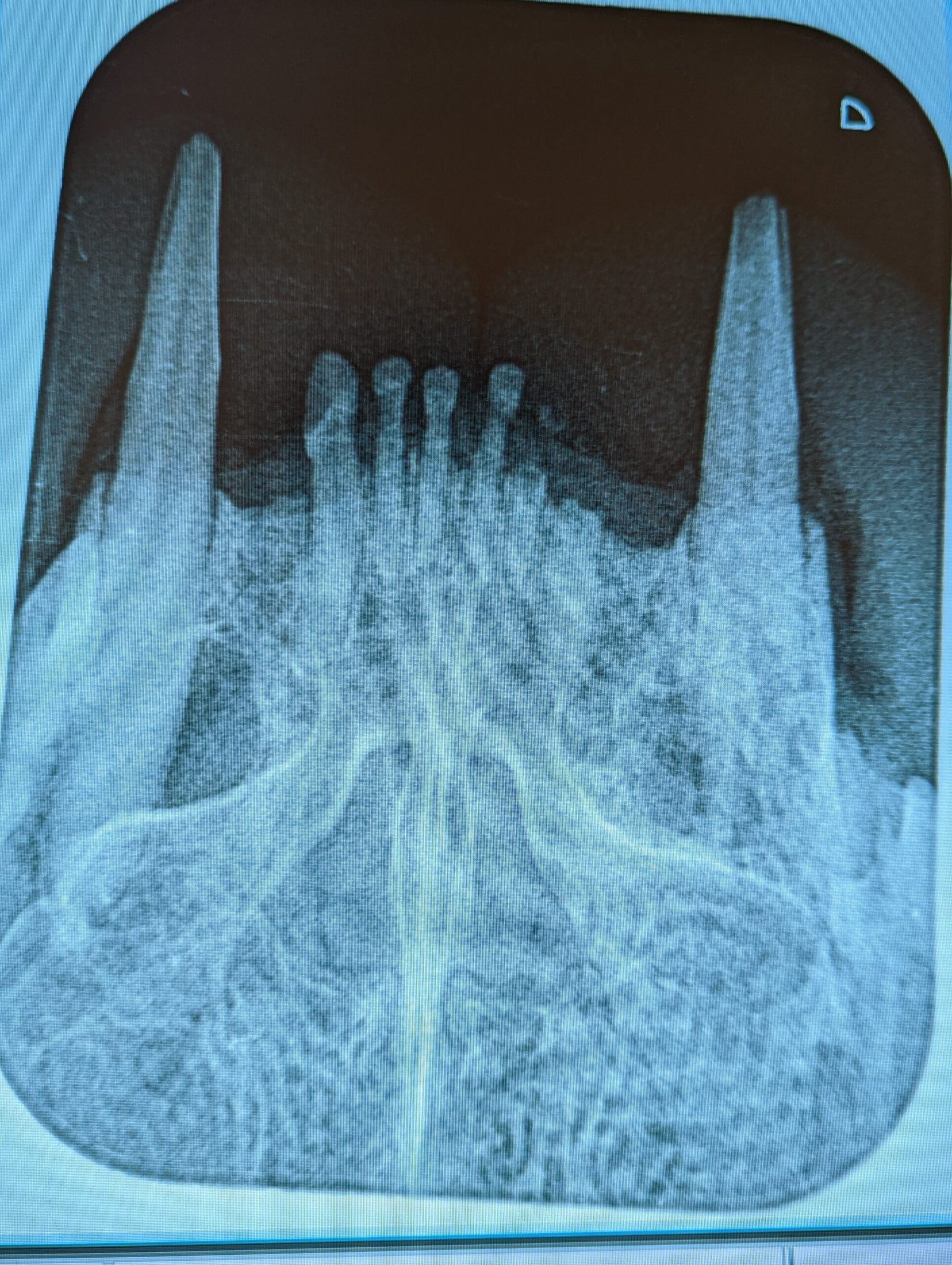
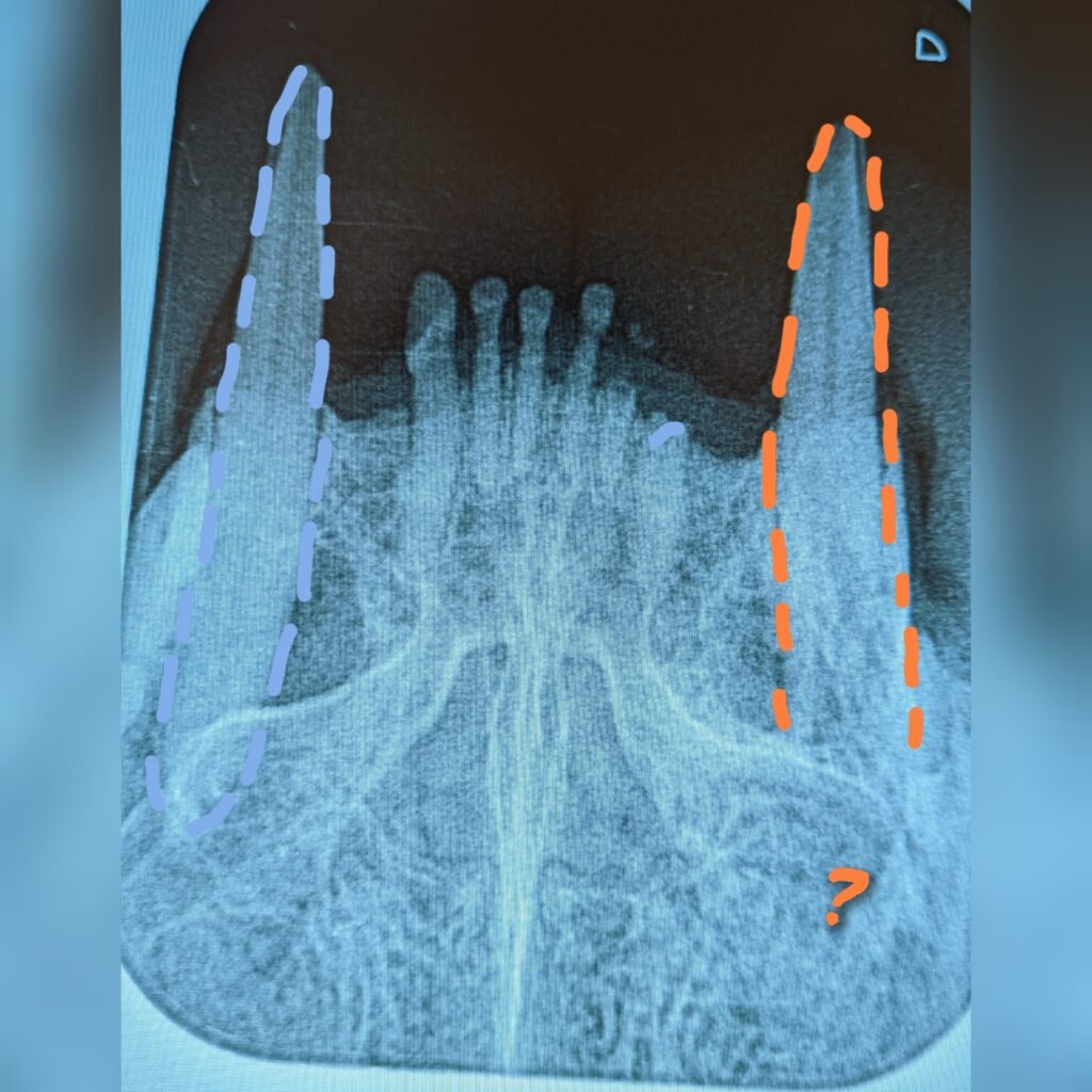
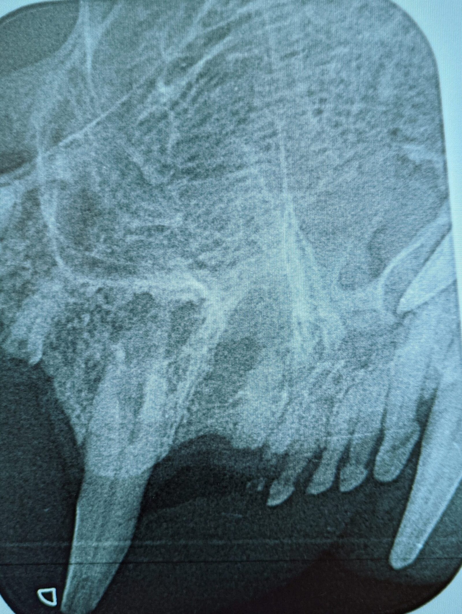
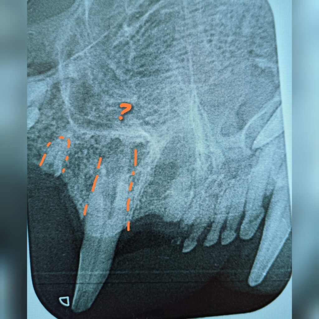
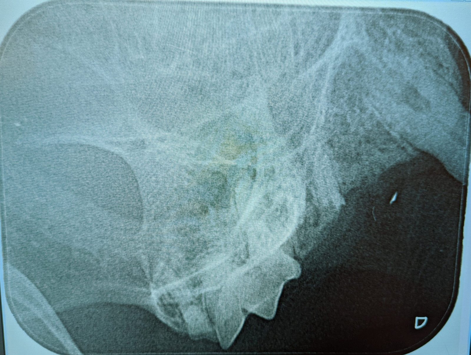
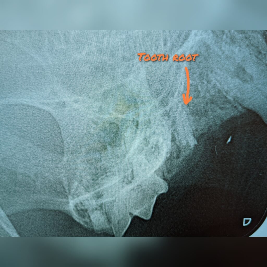
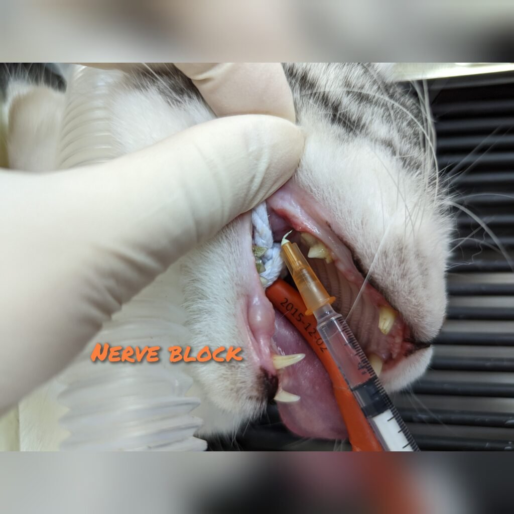
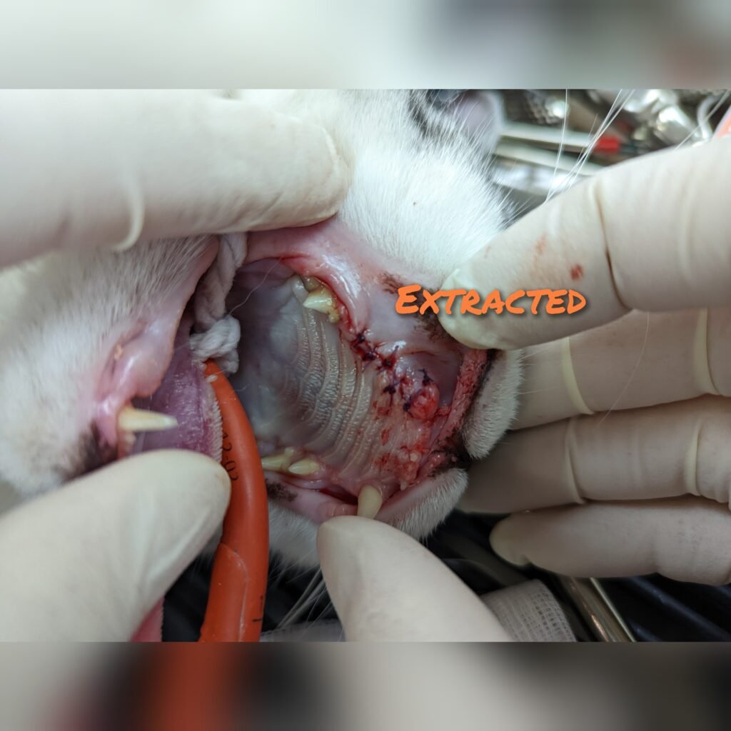
This cat was presented for decrease appetite and having oral pain for a week. He was grossly normal on clinical examination so I decided to examine his teeth under general anesthesia(GA). Grossly, the teeth/gum didn’t look too bad. However the pathology was hidden below the gum and within the root/s🦷. Dental xray is essential to assess any pathology below the gum and the tooth roots. In this case, dental xray revealed 2 broken incisor roots, a diseased canine teeth and a broken premolar tooth root (outlined in orange, pic3/5/7) 😿. Maxillary nerve block (pic 8) was performed to reduce pain and GA risk, and both diseased tooth and roots were surgical removed. (pic 9) 😸
.
Dental disease is like an iceberg 🛳, we only detect about 20-30% dental pathology during clinical examination and another 20-30% under general anesthesia. And the rest of the 50% require dental Xray because the root is as long as the crown.